By: Dr. Dev Batra | 01.30.23

Introduction to Veins
Veins are blood vessels responsible for carrying used blood from your organs and tissues back to your heart. Broadly, we can divide the major veins in our legs into four categories: (1) dermal/hypodermal, (2) superficial, (3) deep, and (4) perforator veins (we won’t discuss perforator veins in detail in this post, but at a high level, these relatively-short vessels “perforate” the fascia and act as a bridge to connect the superficial and deep veins). Altogether there are many routes that blood from the lower extremity can use to return to the heart, but some routes certainly prove more important than others. An obstruction of a primary deep vein or a dysfunctional superficial vein can cause symptoms for patients. In this post, we explore the anatomy of deep and superficial veins.
To go along with our discussion of veins, we have created two cartoon graphics of veins – one healthy, one diseased – at various levels of depth beneath the skin.
Dermal (Cutaneous) and Hypodermal (Subcutaneous) Veins
The dermal veins drain blood from the dermis – one of the most superficial layers of the skin. The hypodermal veins are larger in diameter and reside slightly deeper, but are well-connected to their dermal counterparts. The purpose of these veins is to drain the integumentary system (the skin, hair follicles, nails, and certain glands). Subtle offsets in pressure or flow – sometimes (but not always) due to underlying vein disease (as depicted in Figure 2) – may cause these veins to become bulged and visibly discolored as spider veins or reticular veins. Spider veins are easier to see due to their proximity to the skin surface, and measure up to 1 mm in diameter. Reticular veins may be slightly larger (1-2 mm), less visible, and even serve as deeper “feeder veins” for the spider veins.¹ Regardless of the size, these forms of vein disease are treatable, with the gold standard being sclerotherapy injections which we offer at the Texas Vascular Institute.

Superficial Veins
The superficial veins are larger in diameter, and reside deeper than the dermal and hypodermal categories we just finished discussing – making the term “superficial” somewhat of a misnomer. Two notable veins within this category include the great saphenous vein (GSV) and small saphenous vein (SSV). As Figure 1 depicts, these veins reside within or beneath the layer of fatty (adipose) tissue beneath the skin. A key difference is that the main superficial veins contain one-way valves meant to prevent back-flow (or “reflux”).
The great saphenous vein, for example, travels along the inside of the leg and thigh (about a half-inch beneath the skin surface) until it dives deeper and empties into a deep vein near the groin, eventually leading the blood back to the heart. The GSV’s characteristics vary from patient to patient, but it typically measures 3-5 mm in diameter, and along its length there are typically 5-10 one-way valves.²⁻³ The term varicose vein is used to describe visibly enlarged or tortuous superficial veins. Varicose veins are often a result of the failure of one or more of the one-way valves, resulting in blood flowing backward down the leg (“reflux”), and subsequent clinical signs/symptoms of varicose veins. Treatment of symptomatic varicose veins is offered at the Texas Vascular Institute, and is accomplished by closing off the dysfunctional vein using one of several minimally-invasive techniques.
Deep Veins
The deep veins serve as a central “highway” for blood to make its way back to the heart from the leg. They differ from the superficial veins by virtue of their location, size, quantity, and morphology. Deep veins run in between the various muscles of the leg, where they collect used blood from the deeper tissues (muscles and bone). They also collect blood from the superficial system via deep-superficial junctions (such as the sapheno-femoral junction), as well as perforator veins. When an individual walks, it is the contraction of the calf muscle that drives the flow of blood up the leg through the deep veins. One-way valves are even more abundant in the deep veins than the superficial veins, serving to prevent blood from pooling, and keeping blood flowing in one direction toward the heart.⁵

Below the knee, the average man or woman will have six central deep veins. Moving up the leg at the level of the thigh, the deep veins become fewer in quantity and larger in diameter. Some common deep veins in the thigh area include the femoral vein and the profunda femoris vein, which measure 6-7 mm in diameter.⁴ Higher up and near the groin, the common femoral vein and iliac veins tend to measure greater than 1 cm in diameter. Finally, the common iliac veins from both legs join to form the inferior vena cava (or “IVC”). The IVC measures 1.2-1.7 cm in diameter and travels up through the abdomen to empty into the right side of the heart.⁴
Deep vein thrombosis (DVT) is diagnosed when a blood clot (a “thrombus”) forms in one or more deep vein segments, impeding the flow of blood and causing symptoms such as swelling, redness, and pain. Furthermore, a pulmonary embolism (PE) may result from a DVT that has broken off from the deep vein in the leg and migrated up the IVC through the heart and into the lungs – where it can occlude major blood vessels. The widely-embraced phrase “venous thromboembolism” refers to the combination of DVT and PE. This combination is logical given the circulatory anatomy and the continuous nature of the veins with the heart and lungs. PE can be a life-threatening condition depending on the size of the clot, and the health status of the patient.
About the author
Dr. Dev Batra, M.D. is a vein specialist and founding partner of Texas Vascular Institute. Holding board certifications in radiology and vascular & interventional radiology, he is well-versed in vein issues and has been voted one of D-Magazine’s best doctors in Dallas for three years running.
This blog post was written with research and editorial assistance from OnChart™.
References
[1] Meissner MH. Lower Extremity Venous Anatomy. Seminars in Interventional Radiology. 2005;22(3):147-156.
[2] Cotton LT. Varicose veins: gross anatomy and development. Br J Surg. 1961;48:589-598.
[3] Dan E Spivack et al (2012). Mapping of Superficial Extremity Veins: Normal Diameters and Trends in a Vascular Patient-Population. Ultrasound Med Bio 38(2): 190-4.
[4] BS Hertzberg et al (1997) Sonographic assessment of lower limb vein diameters: implications for the diagnosis and characterization of deep venous thrombosis. American Journal of Roentgenology. 1997;168: 1253-1257.
[5] HM Moore et al (2011) Number and location of venous valves within the popliteal and femoral veins – a review of the literature. J Anat. 2011 Oct; 219(4): 439–443.
Medical Disclaimer
The Materials available in the Texas Vascular Institute blog are for informational and educational purposes only and are not a substitute for the professional judgment of a healthcare professional in diagnosing and treating patients.
Read more blogs
What Is Heat Edema?
Learn what heat edema is, what causes it, and when it could signal a vascular issue. Learn how to manage it and when to seek vascular care.
Why Are My Veins Blue?
Wondering why your veins look blue under your skin? Learn the science behind vein color, how light affects what you see, and what it means for your health.
10 Warning Signs Of Poor Circulation And How To Fix It
Have you ever noticed your feet always feel cold, or your legs cramp up when walking? These could be warning signs of poor circulation, a condition that can impact your daily life and overall health.
WHAT OUR PATIENTS
have to say
Texas Vascular Institute always appreciates feedback from our valued patients. To date, we’re thrilled to have collected 378 reviews with an average rating of 5 out of 5 stars. Please read what others are saying about Texas Vascular Institute below, and as always, we would love to collect your feedback.
Leave a Review
Amazing Practice
I'm very particular with my Healthcare and tend to be cautious with referrals to specialists. This office is amazing from the first point of contact. Their staff are friendly, professional and highly knowledgeable. Then the Dr is just as amazing as his staff, absolutely brilliant. Office manager Jessica has this office running like a well oiled machine and does so with a smile, an air of confidence, kindness and professionalism. Love this practice!!
- Richard G.

Beyond Thankful
Dr Batra and his staff are amazing! We are so grateful to have found him. Everyone is so kind and so caring and Dr Batra explains everything so well and does procedures with excellence. Beyond thankful to be under their care!!!
- Bitsy P.

Gold Standard
This is a gold standard for how a medical practice should be run. I was promptly seen at my scheduled time, my ultrasound was thorough and I received plenty of attention and care from the staff and Dr.Batra.
- Weronika L.
INSURANCE
We accept most major insurance plans. Please contact the medical office for all insurance related questions.


8330 Meadow Rd #100
Dallas, TX 75231
For Appointments: 972-798-4710
General Inquiries: 972-646-8346

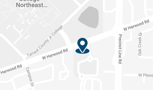
809 West Harwood Rd, Suite 101,
Hurst, TX 76054
For Appointments: 972-798-4710
General Inquiries: 972-646-8346

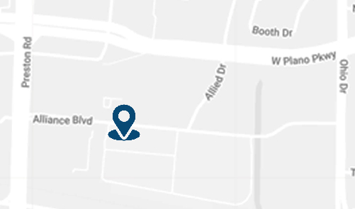
4716 Alliance Blvd Suite #180,
Plano, TX 75093
For Appointments: 972-798-4710
General Inquiries: 972-646-8346

